Comunicación
Nº 056
Comunicación |
S. Ambrosi, M.D., P. Di Zitti, M, M., A. Baiocchini, M. D.
 |
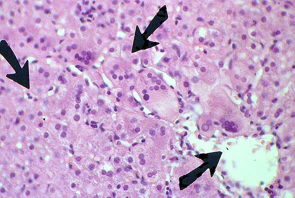
| Fig. 1.- Giant cell transformation (arrows *). Multinucleated hepatocytes in a centrilobular area (H/E x 100). |
 |
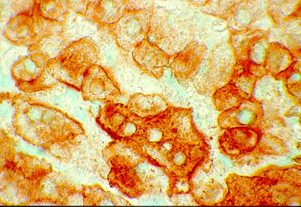
| Fig. 2.-Immunohistochemical labelling with cytokeratin 8,18. Giant cells show the same reactivity of normal hepatocytes (CAM 5.2 x 400). |
 |
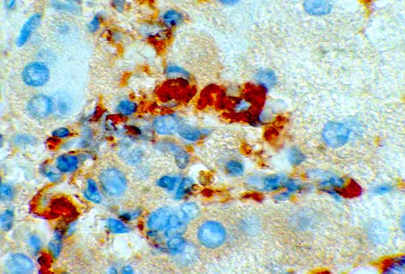
| Fig. 3.- Immunohystochemical labelling with CD 68 showing intense fagocitic activity of giant cells and of Kupffer cells (CD 68 x 400). |