 Virtual Slide # 1. 11-year-old male with bronchiectatic left lung
|
|
Banach Lech*, Mirta Garcia Jardon* |
||
|
11 year-old boy admitted with history of repeated crisis of cough, malaise and fever of two year duration. X-ray images showed complete opacity of the left lung. The patient was previously admitted often with no remission of symptoms. Progression of the pulmonary lesions confined to left lung only made him a suitable candidate for surgery. The patient was referred with clinical diagnoses of tuberculosis and bronchiectasis with recurrent bacterial infections.
12x9x5cm left lung, from total pneumectomy was received at the Department of Pathology. The specimen was fixed in formalin 10% and processed routinely. The appearance of the lung can be seen in the (figure 1). The pleura were opaque, covered by yellowish, fibrinous exudate. Four cm length by 1.5cm in diameter segment of main bronchus was attached to the specimen. Cut section revealed diffuse homogeneous yellowish colour, firm consistency and lack of caseating or suppurative necrosis. No normal parenchyma could be found (figure 1).
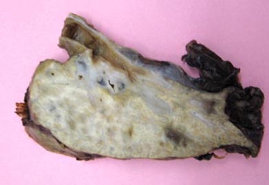 fiogf49gjkf0dFigure 1: Macroscopic appearance of the sectional surface of lung. Notice the yellowish, solid texture, lack of cavitation and/or suppuration.">
Figure 1: Macroscopic appearance of the sectional surface of lung. Notice the yellowish, solid texture, lack of cavitation and/or suppuration. fiogf49gjkf0dFigure 1: Macroscopic appearance of the sectional surface of lung. Notice the yellowish, solid texture, lack of cavitation and/or suppuration.">
Figure 1: Macroscopic appearance of the sectional surface of lung. Notice the yellowish, solid texture, lack of cavitation and/or suppuration.
|
||
|
|
Web mantenido y actualizado por el Servicio de informática uclm. Modificado: 16/06/2015 17:26:32